Your go-to destination to discover practical strategies. No matter where you are in your professional journey, you'll find something valuable here.
360 Neuro Health Blog
The value of the Pupillary Light Reflex (PLR) or Swinging Penlight Test

PURPOSE:
To assess pupil constriction with exposure to direct light.
INDICATIONS:
Assessment of the PLR is useful for identifying a relative afferent pupillary defect (RAPD) due to dysfunction anywhere along the afferent pupillary pathway. The clinician can provide a quick, inexpensive analysis of the patient’s pupillary response to light.
EQUIPMENT:
Penlight with pupil size measurements (millimeters) and distant target. Optional: a distinct target for near-target testing.
TIME REQUIRED:
Less than 2 minutes.
SET-UP:
- Body Position:
- The test is performed with the patient sitting in an upright posture.
- Head Position:
- The test is performed with the head in a neutral position. Avoid forward head posture or head tilt as much as possible.
- Eye(s) Tested:
- Testing is done binocularly (using both eyes).
- Distance of Targets:
- Far Target: 5 feet (1.5 meters) to 10 feet (3 meters)
- Option to add a distinct target for near testing: hold the target 4 inches (10 centimeters) from the bridge of the patient’s nose.
- Distance of Penlight:
- When observing the size and shape of the pupils, hold the penlight horizontally under each eye and use the pupil sizes on the penlight (circles) to measure the size.
- When observing the PLR, hold the penlight 3-6 inches (8-15 centimeters) from the patient’s eyes to direct the light into each eye.
INSTRUCTIONS:
- First, dim the lights in the room if possible.
- Hold the penlight horizontally under the patient’s eye, using positions noted in the “Set-Up” section to identify the patient’s baseline pupil size.
- Instruct the patient: “Look out into the distance while I examine the size and shape of each pupil.” Record the patient’s pupil size using the penlight references (millimeters) and note if pupil sizes are not equal.
- Next, instruct the patient: “Look ahead into the distance [at a specific target] and keep your eyes as wide open as possible. Keep looking ahead while I shine the light back and forth into each eye.” Illuminate the penlight into 1 eye and hold for 3 seconds, then swing to the other eye. Repeat this 2-3 times for each eye.
- If using a near target, instruct the patient: “Hold the target about 4 inches from your face.” Then, add, “Keep looking ahead at the target while I shine the light back and forth into each eye.” Illuminate the penlight into 1 eye and hold for 3 seconds, then swing to the other eye. Repeat this 2-3 times for each eye.
THE VALUE:
The Pupillary Light Reflex (PLR) is an incredibly complex physiological process that involves multiple neurological pathways, providing valuable insights into autonomic, midbrain, and central brain functions. Additionally, a mobile pupil has been found to enhance visual performance by affecting depth of focus and optical abnormalities. This means that studying pupil responses can also have practical applications in improving vision and optimizing visual performance.
CLINICAL TAKEAWAYS:
- If one pupil is consistently larger than the other (anisocoria), it warrants further evaluation. A sudden onset of anisocoria indicates an acute neurological event.
- Pupils constrict due to parasympathetic activity and dilate due to sympathetic activity. Monitoring pupillary changes during position changes helps assess ANS function.
- A sluggish or absent PLR is of significant concern for impaired neurological function, especially if the pupil remains fixed and dilated (mydriasis) during the PLR test.
- If you see a poor response to the PLR test, compare their pupil response to the light versus viewing a near target or a convergence test. Poor response to the PLR test but constriction with viewing a near target (accommodation) is referred to as Light-Near Dissociation, which can occur with neurosyphilis, diabetic neuropathy, lesions in the midbrain, or can occur with a benign condition.
- Bilateral pinpoint, pupils (miosis) may respond to the PLR test, but excessive constriction can occur from opioid use, brainstem dysfunction, neurological conditions, or some medications.
- In concerns of ANS dysfunction, assess the PLR in supine to standing. An abnormal PLR in standing (compared to a normal response in supine) may appear as a sluggish constriction, asymmetry, or a fixed and dilated pupil. These findings suggest ANS dysfunction.
VIDEO DEMONSTRATION:
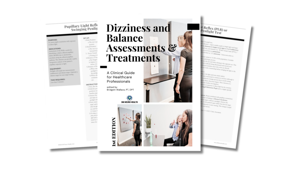
For interpretation and additional considerations, grab your copy of the Dizziness and Balance Assessments & Treatments – A Clinical Guide for Healthcare Professionals | 1st Edition today! Click here to learn more.